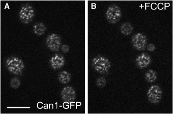
Figure 7.

Filipin stain prevents the release of Can1p from MCC. Cells expressing Can1GFP (strain GYS113) were stained by filipin and observed under agarose. These cells show the characteristic MCC pattern of Can1GFP fluorescence (A). To the same cells immobilized in agarose, 50 μM FCCP was added. The Can1-GFP pattern does not change (B) (compare with Can1-GFP cells depolarized by FCCP without filipin treatment; Figure 5A, right). Bar: 5 μm.
