
Figure 5.
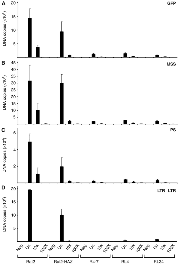
Analysis of the block to virus infection using quantitative RT-PCR. Wt Rat2 cells, the control empty vector Rat2-HAZ line, the control virus-resistant R4-7 line and two of the N-Msn-zeo lines isolated from the screen (RL4 and RL34) were infected with either undiluted (un) or 10-fold serially diluted (10 × , 100 ×) ecotropic MLV-GFP. Uninfected cells were included as a negative control (indicated as neg) for each sample. Total DNA was isolated at 24 h after infection and the amount of viral DNA synthesized in the infected cells was measured by QRT-PCR. Using primers specific to GFP sequences, minus-strand strong stop (MSS) DNA, plus-strand DNA (PS) or LTR–LTR junction, the amount of total linear viral DNA (A), MSS DNA (B), PS DNA (C) or circular viral DNA in the nucleus (D) was determined. Each DNA sample was assayed in duplicate at a minimum of three different time points.