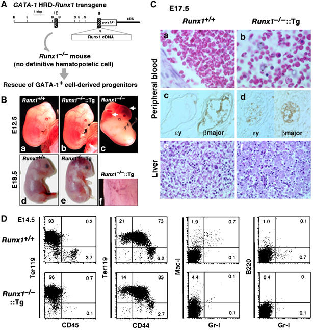
Figure 4.

Rescue of GATA-1+ cell-derived progenitors in Runx1−/− embryos. (A) Strategy for the rescue of GATA-1+ cell-derived progenitors in Runx1−/− embryos. The G1-HRD-Runx1 transgene contains the 3.9-kb sequence 5′ of the IE exon, the IE exon itself, the first intron, and a part of the second exon of the mouse GATA-1 gene in front of Runx1 cDNA. The initiation Met codon in the second exon was replaced by a unique NotI site (shown as N) for subsequent cloning. Restriction enzyme sites are B, BamHI; E, EcoRI; N, NotI; S, SacI. (B) Macroscopic appearance of G1-HRD-Runx1 transgene-rescued embryos. Embryos with each genotype at E12.5 (a–c) and E18.5 (d and e) are shown. A higher magnification picture of the skin of transgene-rescued embryo (f) reveals micro-hemorrhages in the skin. (C) Histological analysis of wild type and Runx1−/−∷Runx1-Tg+ embryos at E17.5. Hematoxylin and eosin staining of peripheral blood from wild type (a) and Runx1−/−∷Runx1-Tg+ (b) embryos are shown. Note that numerous enucleated erythrocytes are present in the Runx1−/−∷Runx1-Tg+ embryos. Panels (c) and (d) show sections stained with anti-βmajor globin antibody or anti-ɛy globin antibody. Hematoxylin and eosin-stained livers of wild type (e) and Runx1−/−∷Runx1-Tg+ (f) embryos. (D) FACS analysis of E14.5 fetal liver cells from wild type and Runx1−/−∷Runx1-Tg+ embryos.
