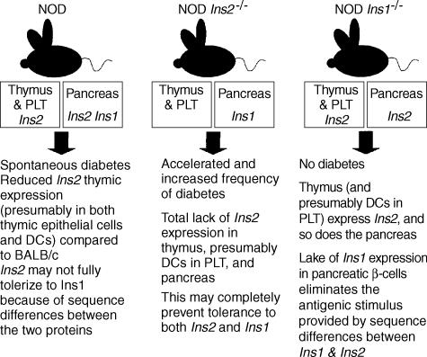
Figure 1.
Illustration of the relationship between the expression of insulin in the thymus and peripheral lymphoid tissues (PLT), mediated by thymic epithelial cells and bone marrow-derived dendritic cells (DCs), and diabetes development in the mouse strains. The figure also highlights the different outcomes associated with the manipulation of the Ins2 and Ins1 genes in relation to their expression in the thymus and PLT, as well as in the pancreas. Please see the text for details. NOD, non-obese diabetic.

