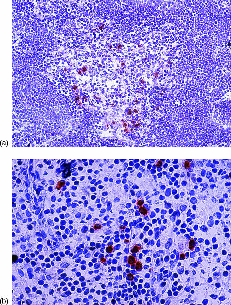
Figure 2.
(a) Proinsulin-positive cells in human thymus, stained on a frozen section using streptavidin–biotin–peroxidase and the AEC substrate (red), counterstained with haematoxylin (×64 magnification). (b) Proinsulin-positive cells in human spleen, stained on a frozen section using streptavidin–biotin–peroxidase and the AEC substrate (red), counterstained with haematoxylin (×128 magnification).

