Figure 5.
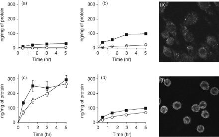
Cellular association time-course experiments of 32P-labelled plasmid DNA (pDNA) in RAW264.7 cells (a), J774A1 cells (b), and resident (c) or elicited (d) macrophages (Mφs); and cellular localization of pDNA in RAW264.7 cells (e) or resident Mφs (f). (a–d) Cells were incubated with 32P-labelled pDNA (0·1 µg/ml) at 37° (closed square) or 4° (open circle). Each point represents the mean ± standard deviation (n = 3). (e and f) Cells were incubated at 4° for 30 min in the presence of 5 µg/ml Texas Red-labelled pDNA. After washing, the cells were warmed to 37° to allow internalization for 15 min. Images represent laser-scanning confocal microscopy sections.

