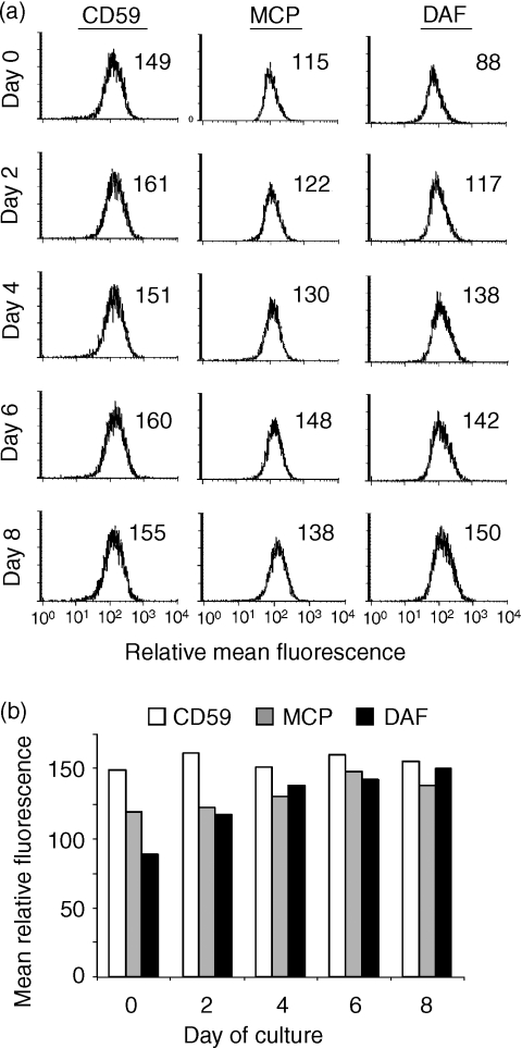
Figure 3.
Effect of dexamethasone on the expression of complement-inhibitory proteins on DU145 cells. Cells were cultured in the absence (or presence) of dexamethasone for the indicated time periods and the expression of complement-inhibitory proteins examined by flow cytometry. (a) All cells were in culture for 8 days, with dexamethasone added at intervals. Day 0 represents culture in the absence of dexamethasone (see ‘Materials and methods’). (b) Graphical summary of flow cytometry data. Staining with a control antibody gave a mean relative fluorescence of <10. The data shown are representative of three separate experiments.

