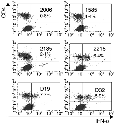
Figure 2.
Intracellular detection of interferon-α (IFN-α) in SWC3+ subsets after stimulation with different CpG-oligonucleotides (ODNs). The SWC3+ cells were enriched by magnetic-activated cell sorter (MACS) separation. After 6 hr of incubation with CpG-ODNs (10 µg/ml), the cells were harvested, stained for cell-surface CD4 and SWC3 antigen, fixed, permeabilized and then stained for IFN-α. The cells were gated for the SWC3+ population to give the dot-plots presented for CD4/IFN-α staining.

