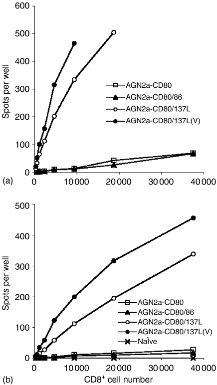
Figure 8.
ELISPOT analysis of CD8 cells in immunized mice. A/J mice were vaccinated s.c with 2 × 10⁶ inactivated AGN2a-CD80, AGN2a-CD80/86, AGN2a-CD80/137L or viable AGN2a-CD80/137L cells twice weekly. At day 5 following the second immunization, CD8+ splenocytes were purified with anti-CD8 immunomagnetic beads from each group, including CD8 splenocytes from naive mice as controls. Decreasing numbers of freshly isolated CD8 T cells (i.e. no in vitro culture) were incubated with 2 × 10⁴ mitomycin C-inactivated cell transfectants of the same origin as those used for vaccination (a) or inactivated wild-type AGN2a cells (b) IFN-γ spots were developed 30–35 hr following incubation, based on the Pharmingen protocol.

