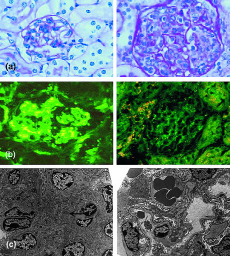
Figure 5.
Glomerulonephritis in C57BL/6 SAP–/– mice. (a) Right panel, typical proliferative glomerulonephritis in SAP–/– mouse showing increased mesangial matrix and marked hypercellularity with mononuclear cells in capillary loops; left panel, normal glomerulus from wild-type mouse (× 250, haematoxylin & eosin stain). (b) Left panel, immunofluorescence stain with anti-mouse IgG showing granular appearance typical of immune complex deposition; right panel, specificity control showing complete abolition of staining after absorption of the primary antibody with mouse IgG (× 250). (c) Left panel, electron micrograph of glomerulus from SAP–/– mouse showing mesangial expansion and immune complex deposition (arrowed); right panel, normal glomerulus from wild-type mouse (× 2000).

