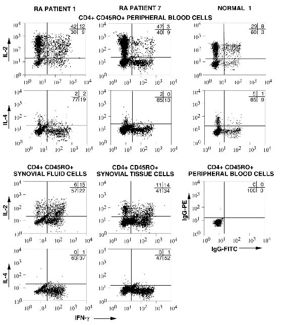
Figure 1.

Representative cytokine profiles of Tm isolated from healthy donors and RA patients. CD4+, CD45RO+ T cells were isolated from the blood and synovial fluid of an RA patient (RA Patient 1), the blood and synovial tissue of an RA patient (RA Patient 7), and the blood of a healthy donor (Normal 1). Intracellular cytokines were assessed for peripheral blood and synovial cells from RA patients or blood of healthy donors. Cells were briefly stimulated for 6 h, fixed, permeabilized and stained with directly labeled control antibodies or anti-IFN-γ-FITC in combination with either PE-labeled anti-IL-2 or PE-labeled anti-IL-4, as indicated on the axes; 104 cells were collected for each analysis. The numbers shown represent the percentages of cells in each quadrant. Control staining with IgG-PE and IgG-FITC was carried out for each sample as shown (Normal 1) and the background was subtracted. Samples were obtained from matching RA blood and synovial fluid (n = 5), matching blood and synovial tissue (n = 3), and normal blood (n = 11). Statistical analysis of all samples evaluated for intracellular cytokines immediately after isolation is shown in Fig. 2.
