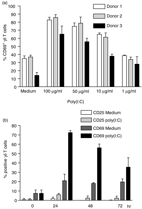
Figure 1.
Partial activation of γδ T cells by polyinosinic-polycytidylic acid [poly(I:C)]. (a) Expression of CD69 on γδ T cells after stimulation of primary peripheral blood mononuclear cells (PBMC) with different concentrations of poly(I:C). Results represent mean values ± standard deviation (SD) of triplicate cultures from three different healthy donors. (b) Percentage of CD69+ and CD25+γδ T cells before (0 hr) and after 24, 48 and 72 hr of primary PBMC culture in the presence of poly(I:C) (100 µg/ml) or medium alone. Results are shown as mean ± SD of triplicate cultures from one representative donor. Similar activation profiles were observed in four additional donors.

