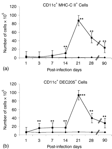
Figure 3.
CD11c+/MHC-CII+ and CD11c+/CD205+ DC increased in the MLN during the first phase of the pulmonary disease. BALB/c mice were infected ITT with M. tuberculosis H37Rv (▪), while control mice were inoculated with sterile saline solution (⋄). MLN cell suspensions from both groups were analysed by flow cytometry on days 1, 3, 7, 14, 21, 28 and 90 postinfection, combining (a) CD11c plus MHC-CII and (b) CD11c plus Dec205. Each group (control and infected) consisted of at least five mice per time point. Results are presented as the mean ± standard error of three different experiments. Statistically significant differences are indicated by asterisks (*P < 0·05; **P < 0·01; ***P < 0·001).

