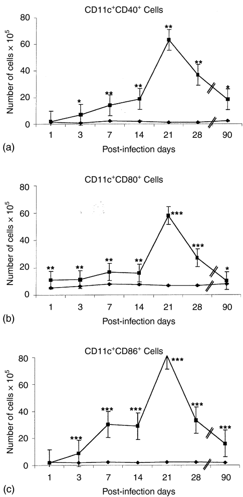
Figure 4.
Expression of costimulatory molecules (CD40, CD80 and CD86) in CD11c+ DC during the course of pulmonary disease. BALB/c mice were infected ITT with M. tuberculosis H37Rv (▪) while control mice were inoculated with sterile saline solution (⋄). MLN cell suspensions from both groups were analysed by flow cytometry on days 1, 3, 7, 14, 21, 28 and 90 postinfection, combining (a) CD11c plus CD40 (b) CD11c plus CD80, and (c) CD11c plus CD86. The mean result ± standard error of three separate experiments is shown, significant differences are indicated by asterisks (*P < 0·05; **P < 0·01; ***P < 0·001).

