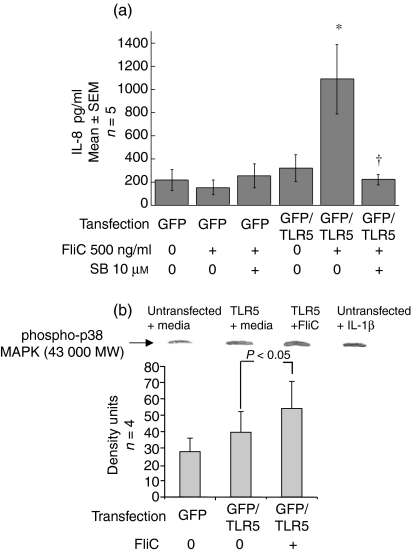
Figure 4.
(a) Inhibition of FliC-EAEC-induced IL-8 secretion from TLR5-transfected HEp-(a) 2 cells. Untransfected and TLR5-transfected HEp-2 cells were preincubated with DMSO or SB-203 580 (10 µm) for 1 hr and then treated with FliC-EAEC (500 ng/ml) for 3 hr. Transfection efficiency was 70–80% as verified by fluorescent microscopy to detect GFP. IL-8 was measured by ELISA. Overall P < 0·005 by anova. *P < 0·05 vs. untransfected HEp-2 cells treated with FliC and vs. TLR5-transfected cells with media alone. †P < 0·05 vs. TLR5-transfected cells treated with FliC alone. (b) Activation of p38 MAP kinase in TLR5-transfected HEp-2 cells exposed to FliC-EAEC. Transiently transfected HEp-2 cells were incubated with media alone or FliC-EAEC (500 ng/ml) for 1 hr and were analysed by Western blots with rabbit anti-phospho-p38 MAP kinase antibody. Controls included untransfected HEp-2 cells treated with media alone or with IL-1β (10 ng/ml). Equal protein loading was verified by India ink staining. Bands were compared by densitometry and analysed by t-test.

