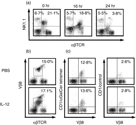
Figure 1.
NK1.1+T cells in the liver became undetectable after IL-12 injection into mice, whereas Vβ8+T cells with lower TCR and CD1d/α-GalCer tetramer binding Vβ8+T cells remain detectable. (a) NK1.1 expression of liver αβT cells. The middle-aged (25-week-old) mice were injected intraperitoneally with IL-12 and livers were obtained at the indicated times after IL-12 injections. Liver MNC were stained and analysed. Numbers of upper quadrants represent percentage of NK cells and NK1.1+T cells in the liver MNC. (b) Vβ8 expression of liver αβT cells. Numbers within left panels represent percentage ofVβ8T cells with intermediate TCR in whole liver MNC (indicated by squares). (c) CD1/α-GalCer tetramer binding Vβ8+T cells. Livers were obtained from middle-aged mice 24 hr after IL-12 injection and then the liver MNC were stained and analysed.

