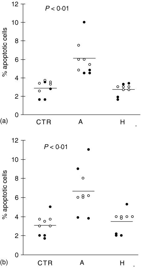
Figure 1.
Basal values of monocyte and T lymphocyte apoptosis in VL patients. Blood samples were collected from healthy controls (CTR) and 9 VL patients at the time of acute disease (A) and after healing (H) (paired samples) and, hence, evaluated for spontaneous basal apoptosis by FACScan cytometer analysis using propidium iodide (PI) and anti-CD14 mAb to evaluate monocytes (a), and anti-CD3 to estimate lymphocytes (b). Each circle represents a subject. Open circles indicate those patients that were followed through in later studies and showed in other figures and tables. Bars represent the mean value in each group. P < 0·01: significant differences from the mean value of the same patients studied in healed phase of disease and from healthy controls. Basal percentage of monocytes: (a) 16 ± 5; (H) 15 ± 2; (CTR) 14 ± 4. Basal percentage of lymphocytes: (a) 78 ± 10; (H) 70 ± 5; (CTR) 77 ± 9.

