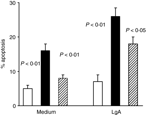
Figure 3.
CD3+ T-cell apoptosis in VL patients cultured in vitro with or without specific antigen. Frozen PBMC from five VL patients, studied in acute and healed phases of disease, and from five healthy controls were cultured for 24 hr in medium alone and in the presence of LAg. Cells were stained with anti-CD3+ mAb and PI and percentage of apoptotic cells evaluated by double staining. Histograms represent median values of the percentage of apoptotic monocytes among CD3+ cells. Healthy controls (white columns); acute VL patients (black columns); healed subjects (shaded columns). Significant differences between acute VL patients and other two groups are shown. The activation with LAg increased significantly (P < 0·01) PCD in acute and healed patients compared with that detected in unstimulated cultures.

