Figure 2.
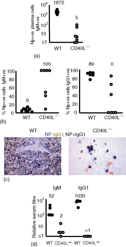
The extrafollicular antibody response is severely impaired but not completely ablated in the absence of CD154. Mice were immunized with NP-CGG and killed B. pertussis and responses assessed 7 days after immunization. Closed and open circles, respectively, represent individual lymph node responses from wild-type and CD154-deficient mice. (a) Numbers of NP-specific plasma cells per section recruited into the extrafollicular response. In five of eight non-immunized CD154-deficient lymph node sections no IgM or IgG1 plasma cells were observed and in all lymph node sections no NP-specific cells were detected. The other three lymph node sections contained 1, 2 and 5 IgG1 plasma cells (data not shown). (b) The proportion of the NP-specific plasma cells shown in (a) that were non-switched or switched to IgG1 7 days after immunization. In CD154-deficient mice, the majority of NP-specific extrafollicular cells are unswitched (left hand panel). In contrast, most plasma cells in wild-type mice have switched to IgG1 (right hand panel); some NP-specific IgG1 cells are detectable. (c) Immunohistology showing the clear IgG1 dominance of the NP-response in wild-type mice (left hand panel) and the clear detection of reduced numbers of IgG1-producing NP-specific cells in some CD154-deficient mice (right-hand panel). (d) NP-specific serum titres, non-immunized mice had antibody titres < 1 (data not shown). Median values for each of the groups are included above the data points. Data are representative of at least two repeat experiments.

