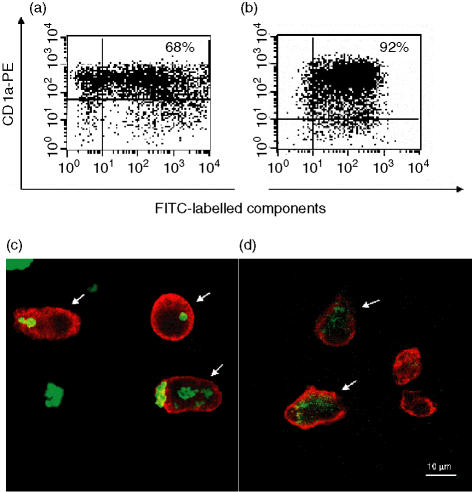
Figure 1.
Uptake of carbohydrate-based particle (CBP)–rFel d 1 complexes, or rFel d 1 alone, by immature monocyte-derived dendritic cells (iMDDCs). MDDCs were incubated for 24 hr with fluorescein isothiocyanate (FITC)-labelled CBP–rFel d 1 or rFel d 1, then stained with anti-CD1a–phycoerythrin (PE) and analysed by flow cytometry (a and b) and confocal laser-scanning microscopy (CLSM) (c and d). More than 60% of the CD1a+ MDDCs were positive for FITC-labelled CBP–rFel d 1 (a), and more than 90% of the CD1a+ MDDCs were positive for FITC-labelled rFel d 1 (b). The percentage of double-positive cells is indicated in the upper right-hand corner. The results presented here are from one of five independent experiments. Multiple Z-series' images obtained by CLSM revealed that the CBP–rFel d 1 (c) and rFel d 1 (d) had been internalized by the MDDCs. Cells sectioned through their centre are indicated with arrows. These images have been optically merged, with red representing Alexa 546-conjugated antibodies detecting CD1a, and green representing FITC-labelled CBP–rFel d 1 or rFel d 1. The scale bar in (d) is also valid for (c). Here, the results from one of four experiments are shown.

