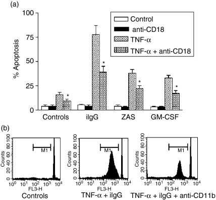
Figure 1.
Promotion of neutrophil apoptosis by tumour necrosis factor-α (TNF-α) involves a Mac-1-dependent pathway. Neutrophils (2·5 × 106/ml) were cultured for 30 min at 4° alone or in the presence of saturating concentrations of blocking monoclonal antibodies (mAbs) directed to CD18 [TS 1/18 F(ab′)2] (a) or CD11b (Mo1) (b). Then, cells were cultured in the presence or absence of TNF-α (10 ng/ml) for 1–2 min at 37° and treated with immobilized immunoglobulin G (iIgG), zymosan-activated serum (ZAS) (1/10) or granulocyte–macrophage colony-stimulating factor (GM-CSF) (50 ng/ml) (a) or iIgG (b). After 3 hr, the percentage of apoptotic cells was determined by fluorescence microscopy (a) or by flow cytometry (b), as described in the Materials and Methods. (a) Results are expressed as the mean ± standard error of the mean (SEM) of five or six experiments. *Statistical significance (P < 0·01) compared to neutrophils cultured with TNF-α in the absence of anti-CD18-blocking antibodies. (b) Histograms of a representative experiment (n = 4) in which M1 represents the fraction of nuclei with hypodiploid DNA content. Sub-M1 events represent cell debris.

