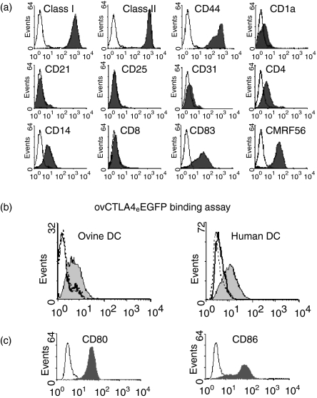
Figure 2.
Phenotypic analyses of ovine lymphatic dendritic cells and the binding activity ovCTLA4eEGFP by flow cytometry (a) Ovine DC obtained by pseudo-afferent cannulation of the prefemoral lymph node were stained with control isotype antibodies (open histograms) and a panel of cell-surface antigen markers (filled histograms). Each histogram is representative of at least three different experiments. (b) Human or ovine DC were incubated with ovCTLA4eEGFP derived from recombinant adenovirus modified fibroblasts. The bound ovCTLA4eEGFP was detected with an anti-GFP mAb as shown in the shaded histogram and the negative-isotype matched mAb (X63) represented by the solid line. The dotted line represents blockade of ovCTLA4eEGFP binding with CTLA4-Ig. (c)Human DC were stained with anti-CD80 and -CD86 mAbs to confirm the presence of the CTLA-4 receptors. Flow cytometric analysis of CD80 and CD86 staining is represented by the shaded histogram with X63 represented by the unshaded histogram.

