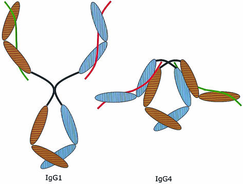
Figure 5.
Proposed model of the overall structure of IgG4 (on the right) compared to IgG1 (on the left). The brown and blue ovals are the domains of the two heavy chains. The red and green lines represent the two light chains. Note the compact structure due to the proposed interaction between the CH1 domain and the trans-CH2 domain. This model does not reflect the difference in structure between inter- and intrachain disulphide bridges in the IgG4 hinge. IgG4 with intrachain bridges is supposed to have an even more compact structure than IgG4 with interchain bridges, but in both cases the CH1 domains interact with the CH2 domains. Moreover, this model depicts IgG4 as a symmetric molecule, as it would be at the time of excretion by the plasmacell. After excretion, the scrambling process illustrated in Fig. 6 will occur.

