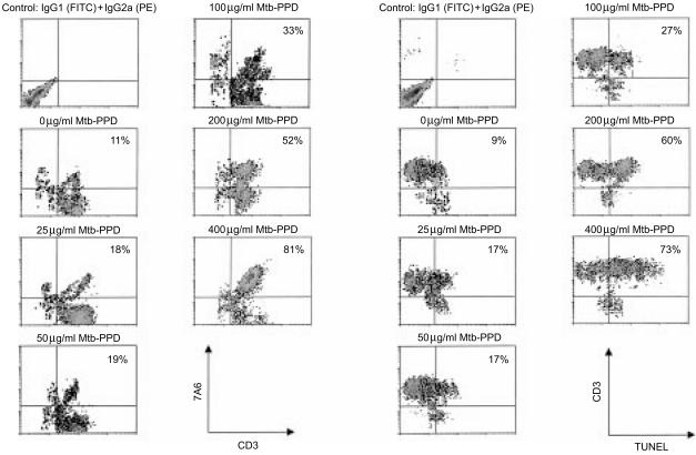
Figure 4.
Apoptosis of Mtb-PPD-specific T lymphocytes following reactivation. Human lymphocytes were cultured with autologous DC and Mtb-PPD (autologous MLC) for 6 days. Following separation from MLC sensitized T lymphocytes were reactivated by different concentrations of Mtb-PPD. The apoptotic rate of reactivated lymphocytes were determined by flow cytometric analysis of the apoptosis-associated mitochondrial membrane protein 7A6 (left panel) and TUNEL (right panel). Two-colour dot plots of a representative experiment with lymphocytes from a donor indicate that the apoptotic rate of reactivated CD3+ lymphocytes is positively correlated with increasing Mtb-PPD concentrations.

