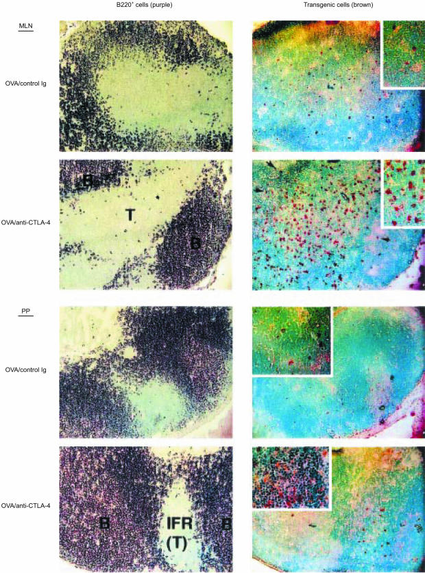
Figure 3.
Immunohistochemical analysis of mesenteric lymph nodes and Peyer's patches. BALB/c mice were treated and killed as described in the legend to Fig. 1b. Serial tissue sections were stained with either biotinylated anti-B220 monoclonal antibody (mAb) (to identify the B-cell-rich follicles) or biotinylated KJ1-26 mAb (to identify T-cell receptor [TCR] transgenic T cells), as described in the Materials and methods. B220+ cells are shown in purple and KJ1-26+ cells are shown in brown. Original magnifications: ×100. B, B-cell follicle; IFR (T), interfollicular region (T-cell zone); MLN, mesenteric lymph node; PP, Peyer's patch; T, T-cell zone. Arrows indicate transgenic T cells in the B-cell follicles. Inserts represent high-power magnifications (× 200) of the same tissue sections. The sections shown are representative of two independent experiments.

