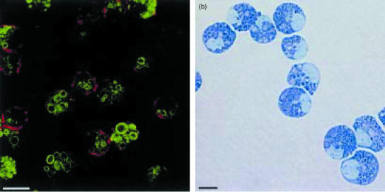
Figure 2.
Confocal image (a) of 14-day-old-mouse bone marrow cultures demonstrating the presence of mature mast cells with abundant granules containing mouse mast cell proteinase-1 (green fluorescence) and expressing the integrin αE (red fluorescence) on their surface membranes. A light micrograph (b) of the mouse bone marrow mast cells (mBMMC) stained with Leishman's shows that they are mature, heavily granulated cells. The cells were grown in medium containing recombinant mouse interleukin (IL)-3, IL-9 and stem cell factor (SCF) supplemented with recombinant human transforming growth factor-β1 (TGF-β1), as described in detail by Miller et al.134 (Horizontal bars represent 10 µm.)

