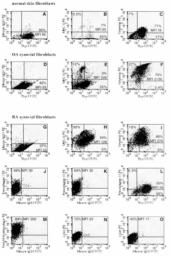
Figure 6.

Phenotype of isolated primary-culture RA-SFB (lower panels) in comparison with primary-culture normal skin-FB (upper panel) and isolated primary-culture OA-SFB. The double-staining experiments were performed with the anti-Thy-1 mAb AS02. (A)-(C) The expression of Thy-1 (A), MHC-II/Thy-1 (B) (double-staining), and vimentin/Thy-1 (C) (double-staining) in normal skin-FB; (D)-(F) the expression of Thy-1 (D), MHC-II/Thy-1 (E) (double-staining), and vimentin/Thy-1 (F) (double-staining) in OA-SFB; (G)-(I) the expression of the antigens Thy-1 (G), MHC-II/Thy-1 (H) (double-staining), vimentin/Thy-1 (I) (double-staining) on RA-SFB; (J)-(L) the expression of the cytoplasmic antigens procollagen I (J) and procollagen III/Thy-1 (K) (single-staining for procollagen III) and (L) (double-staining) in RA-SFB. Expression of prolyl-4-hydroxylase (M) and the proto-oncogenes c-Fos (N), and c-Jun (O) in RA-SFB is also shown. See Results and Table 4 for mean values and statistical comparison among the different FB types. PE, phycoerythrine; FITC, fluoresceine isothiocyanate.
