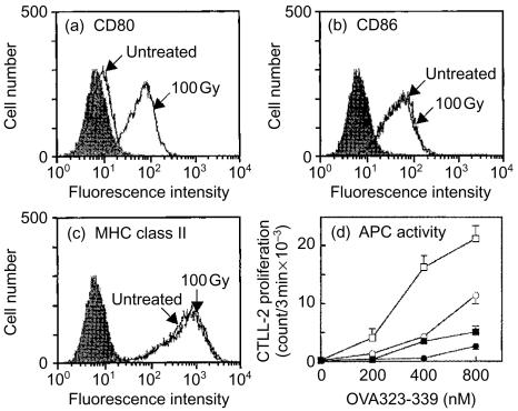
Figure 1.
Irradiation-induced up-regulation of CD80 expression on A20-HL cells. A20-HL cells were X-irradiated with 100 Gy and incubated for 24 hr. Expression of CD80, CD86, and MHC class II molecules was analysed on a flow cytometer. The cells were treated with mAb 16-10A1 (a; anti-CD80, hamster IgG), GL-1 (b; anti-CD86, rat IgG2a), or M5/114 (c; anti-I-Ad, Ed, rat IgG2b), followed by staining with an appropriate second antibody. Filled histograms were controls treated with hamster IgG (a), R34-95 mAb (b; rat IgG2a), or GK1.5 mAb (c; rat IgG2b), as controls. In (d), A20-HL cells were irradiated (squares) or untreated (circles), incubated, and fixed with paraformaldehyde. These fixed cells were incubated for 20 hr with 42-6A T cells and OVA323–339 peptide at indicated doses in the presence of 20 µg/ml anti-CD80 mAb, 16-10A1 (closed symbol), or hamster IgG as a control (open symbol). The culture supernatant (50 µl/well) was examined for IL-2 activity using CTLL-2 proliferation.

