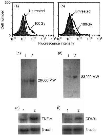
Figure 3.
Irradiation induces TNF-α and CD40L expression. A20-HL cells were irradiated with 100 Gy, and incubated for 24 hr. They were analysed for the cell surface expression of TNF-α (a) and CD40L (b), using anti-TNF-α mAb, G281-2626 (rat IgG1), and anti-CD40L mAb, MR-1 (hamster IgG), respectively, with appropriate second Abs. Monoclonal rat IgG1 (R3-34) (a) and hamster IgG (b) were used as control Abs (filled histograms). Cellular TNF-α(c) and CD40L (d)were analysed by Western blot. Cell lysates were prepared from A20-HL cells untreated (lane 1) and from those 100 Gy-irradiated (lane 2). Blots were probed with a biotinylated anti-TNF-α (c) or anti-CD40 (d) mAb, and visualized. Northern blot analysis of TNF-α (e) and CD40L (f) mRNA expression was also carried out. Cellular RNA was obtained from A20-HL cells untreated (lane 1) or 100 Gy-irradiated (lane 2), electrophoresed, and hybridized with 32P-labelled TNF-α (e) or CD40L cDNA (f) probe. As controls, the membranes were re-hybridized with β-actin cDNA.

