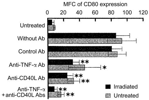
Figure 4.
Marked decrease in the irradiation-induced up-regulation of CD80 expression by anti-TNF-α and anti-CD40L mAbs. Irradiated A20-HL cells were cocultured with those untreated but BCECF-labelled in the presence of 20 µg/ml anti-TNF-α mAb (G281-2626, rat IgG1), 20 µg/ml anti-CD40L (MR-1, hamster IgG), or combination of these mAbs. Rat mAb, R3-34 (rat IgG1), and hamster IgG were used as controls. Then, BCECF+ and BCECF− cells were analysed for the expression of CD80, as described in the legend to Fig. 1. The results were expressed as MFC of CD80 expression. Each bar represents the mean with ±SD from six experiments. An asterisk indicates statistical significance relative to the cocultures (*P<0·05, **P<0·01).

