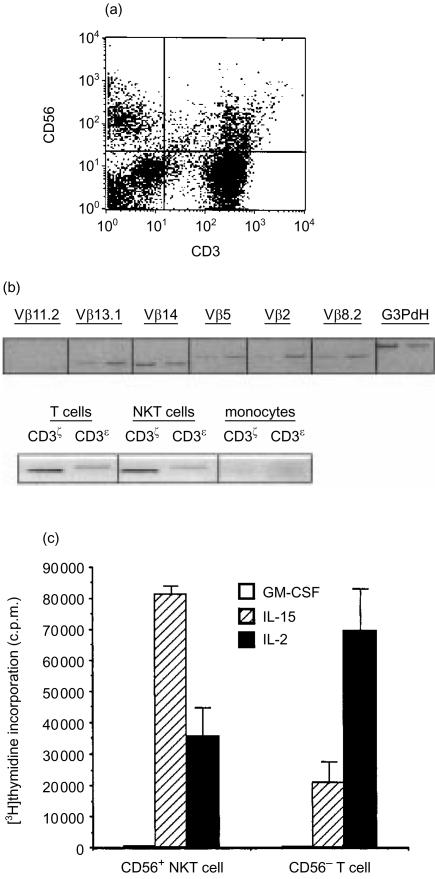
Figure 1.
Peripheral blood mononuclear cells coexpressing NK-and T-cell markers. (a) Coexpression of CD56 and CD3 delineates a distinct mononuclear cell population by flow cytometry. phycoerythrin (PE)-conjugated anti-CD56 and FITC-conjugated anti-CD3 monoclonal antibodies. (b) CD56−NKT express non-biased and rearranged T-cell receptors, and CD3 molecules. For T-cell receptor analysis, the first band is from CD56+ T-cell RNA and the second band is from CD56− NKT cell RNA. Gene-specific Vb oligonucleotide primers combined with a Cβ primer were used for PCR analysis of T-cell receptors. (c) Proliferation of freshly isolated NKT or T cells (2×10⁵/well) incubated with IL-2, IL-15, or GM-CSF for 6 days. Mean of triplicate [3H]thymidine incor-poration ±SEM. Representative experiment of three performed.

