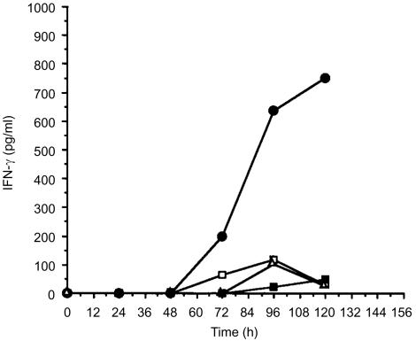
Figure 6.
T-cell recognition of processed antigen displayed by GM-CSF-activated NKT cells. The HC polypeptide of tetanus toxin and GM-CSF were added to freshly isolated NKT cells (1·5×105/well). Autologous T cells (3×10⁵/well) were added at timed intervals from the start of culture and T-cell stimulation was assessed by release of IFN-γ. NKT and T cells without antigen (□); NKT and T cells cultured with antigen (25 µg/ml) (•); NKT and T cells cultured with antigen (25 µg/ml) but without GM-CSF (▪); T cells only with GM-CSF (▵).

