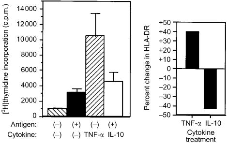
Figure 7.
Differential effect of TNF-α and IL-10 on NKT cells activated with GM-CSF. NKT cells were cultured with GM-CSF for 48 hr followed by the addition of TNF-α or IL-10. Naïve T-cell responses to HC polypeptide of tetanus toxin (10 µg/ml), presented by NKT cells activated with GM-CSF and treated with IL-10 or TNF-α (P<0·005, compared to controls or IL-10 treatment). Data (mean± SD) are representative of three determinations. Cell surface HLA-DR. NKT cells were labelled with FITC-conjugated anti-HLA-DR mAb and cell-associated fluorescence was measured by flow cytometry 3 days after addition of TNF-α or IL-10. Data are plotted as per cent change in cell-surface HLA-DR in comparison to NKT cells treated only with GM-CSF. Results are representative of three determinations.

