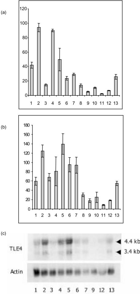
Figure 3.
B-cell-restricted RNA expression of QD and TLE4 transcripts. (a) and (b) Semiquantitative reverse transcription–polymerase chain reaction (RT–PCR) expression of QD (a) and TLE4 (b) transcripts after actin calibration. RT–PCR was performed using a 5′ oligonucleotide in the Q domain common to the TLE4 and QD transcript, and a 3′ oligonucleotide in the 3′-untranslated region of the QD cDNA or in the TLE4 GP domain (see Fig. 1b), leading to PCR products of 515 bp and 238 bp, respectively. The following cell lines were tested: pro-B, lanes 1 (BV173), 2 (JEA2) and 3 (Reh); pre-B, lanes 4 (LAZ) and 5 (Nalm6); B, lanes 6 (Daudi), 7 (Namalwa) and 8 (JY); monocytes, lanes 9 (HL60), 10 (THP1) and 11 (U937); T, lanes 12 (Hsb2) and 13 (Jurkat). Average values of the relative quantity of PCR transcripts for three separate experiments are shown. (c) A Northern blot from the cell lines indicated above was hybridized with the TLE4 (see Fig. 1b) and the actin probes, successively.

