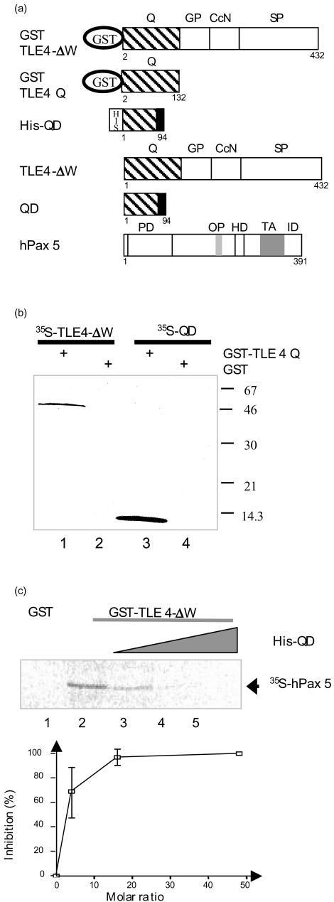
Figure 5.
Analysis of QD, TLE4 and Pax5 protein interactions. (a) Schematic diagram of TLE4, QD and Pax5 fusion proteins. The different domains of each protein are indicated (PD, paired domain; OP, octapeptide; HD, partial homeodomain; TA, transactivation region; ID, inhibitory domain; also see Fig. 1 legend). (b) The QD recombinant protein interacts with the Q domain of TLE4. GST pull-down assays were used to analyse the interactions between in vitro-translated 35S-labelled TLE4-ΔW (lanes 1 and 2) and 35S-labelled QD proteins (lanes 3 and 4) and GST or GST-TLE4Q proteins bound to glutathione-sepharose. (c) The QD recombinant protein inhibits the TLE4/Pax5 interactions. Upper panel: GST pull-down assays were performed with 35S-labelled hPax5 protein and GST (lane 1), GST-TLE4-ΔW (lanes 2–5) bound to glutathione-sepharose. The GST-TLE4-ΔW proteins were incubated with an increasing amount of His-QD proteins (0·5, 1 and 3 µg, lanes 3, 4 and 5, respectively). Lower panel: for three independent experiments the percentage of inhibition was calculated for each QD/TLE4 molar ratio.

