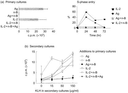
Figure 1.
n-Butyrate does not induce anergy in interleukin-2 (IL-2)-stimulated T helper 1 (Th1) cells. (a) Th1 cells (clone D9) were incubated for 48 hr in primary culture with antigen (Ag) alone, n-butyrate alone, Ag+n-butyrate, recombinant (r)IL-2 alone, rIL-2+n-butyrate, or rIL-2+n-butyrate+Ag. In addition, Th1 cells were stimulated with IL-2 or Ag + n-butyrate for 24, 48, or 72 hr. Following isolation from the primary cultures, the Th1 cells were fixed in 70% ethanol at 4° overnight. Th1 cells were then washed in phosphate-buffered saline (PBS), resuspended in RNAse (1 mg/ml) and propidium iodide (50 µg/ml), incubated for 20 min at room temperature in the dark, and analysed for their DNA content by flow cytometry. (b) Th1 cells were then isolated from the primary cultures and stimulated in secondary cultures with Ag for 48 hr. [3H]Thymidine ([3H]TdR) uptake by the Th1 cells was assessed and is presented as counts per minute (c.p.m.)±SD from a representative experiment. *The responses generated by the Th1 cells pretreated with Ag+n-butyrate or with rIL-2+Ag+n-butyrate and then restimulated with 50 or 150 µg/ml of keyhole limpet haemocyanin (KLH) in secondary cultures were determined by the Student's t-test to be statistically different at a P-value of 0·05 from their non-Ag control values. This experiment has been repeated twice with similar results.

