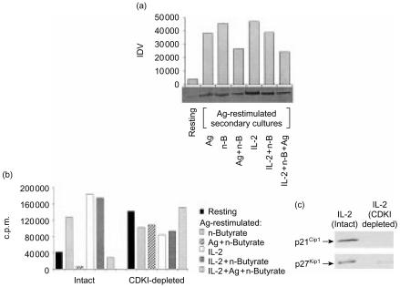
Figure 6.
(a) Endogenous retinoblastoma protein (pRb) levels in anergic and non-anergic T helper 1 (Th1) cells following restimulation with antigen (Ag). Lysates were prepared from resting Th1 cells, or from Th1 cells exposed in primary cultures to Ag alone, n-butyrate alone, Ag+n-butyrate, interleukin-2 (IL-2) alone, IL-2+n-butyrate, Ag+n-butyrate, or IL-2+Ag+n-butyrate, and which had been restimulated with Ag for 24 hr. These lysates were immunoblotted with anti-pRb Ab. Densitometric analysis was performed, and the results are presented as integrated density value (IDV). (b) Activity of cyclin-dependent kinase inhibitors (CDKIs) in anergic and non-anergic Th1 cells following restimulation with Ag. Lysates were prepared from resting Th1 cells, or from Th1 cells exposed in primary cultures to n-butyrate alone, Ag+n-butyrate, IL-2+n-butyrate, or IL-2+Ag+n-butyrate, and then recultured with Ag for 24 hr. The Th1 cell lysates, either intact or depleted of both p21Cip1 and p27Kip1 by immunoprecipitation, were mixed in equal protein amounts with cdk2 immunoprecipitated from lysates of asynchronous EL4 cells for 30 min, and then tested for activity in an H1 kinase assay. The results are presented as total counts per minute (c.p.m.) minus the background c.p.m. measured in samples containing immunoprecipitated cdk2 but no substrate. This experiment was repeated with similar results obtained. (c) Representative experiment demonstrating the immunodepletion of p27Kip1 and p21Cip1 from the Th1 cell lysates (treated with IL-2) confirmed by Western blotting.

