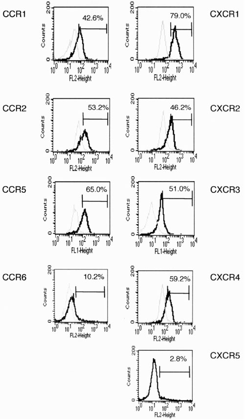
Figure 1.

Chemokine receptor expression by peripheral blood monocytes. Peripheral blood SRBC rosette negative cells (1 × 10⁵) were stained with anti-CD14 mAb and various chemokine receptor mAbs, and were analyzed by flow cytometry. CD14+ cells were gated, and chemokine receptor expression by the CD14+ monocytes is shown (solid line). Dotted lines show staining by isotype-matched control mAb. Percentage of positive cells is also shown in the histograms. Staining was from one of nine experiments. FL-1 height, FITC fluorescence; FL-2 height, PE fluorescence.
