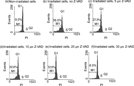
Figure 3.
Detection of apoptotic cells with fractional DNA content based on cellular DNA content analysis. Non-irradiated (a) and gamma-irradiated (b) feeder cells were cultured for 48 hr. Some of the latter cells were cultured with 5 µm (c), 10 µm (d), 20 µm (e), or 30 µm (f) caspase inhibitor Z-Val-Ala-Asp-CH2F (Z-VAD.fmk) from the beginning of culture. The cells were then fixed in 70% ethanol, permeabilized and stained with propidium iodide (PI), and their fluorescence was measured by flow cytometry. The population of early and late apoptotic cells is represented by the M1 fraction.

