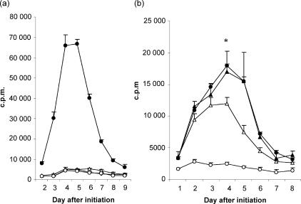
Figure 6.
(a) Peripheral blood mononuclear cells (PBMC) from normal individuals were initially stimulated with 50 µg/ml bovine nucleohistone (NH) (closed circles), 10 µg/ml hen-egg lysozyme (HEL) (open circles), 10 µg/ml bovine thyroglobulin (Tg) (open triangles) or medium alone (open diamonds), for 11 days. T-cell proliferation assays were carried out with these cells stimulated with an equivalent number of irradiated autologous feeder cells. (b) Day-11 NH-primed cells were challenged with feeder cells (closed circles) and day-11 Tg-primed cells were stimulated with 10 µg/ml bovine Tg alone (open circles), feeder cells alone (open triangles), or Tg plus feeder cells (closed triangles). Each point represents the mean value of triplicate wells (1 × 10⁵ primed cells and 1 × 10⁵ feeder cells per well), and error bars indicate the standard deviation (SD). The data are representative of five experiments. *P < 0·05 comparing Tg-primed cells stimulated with feeder cells alone or with Tg plus feeder cells. c.p.m., counts per minute.

