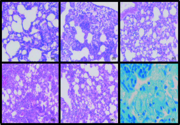
Figure 5.
Morphology of the lungs of mice during active, latent and reactivated tuberculosis infection. Lungs were examined at 2 weeks (a), 10 weeks (b) and (c), and 55 weeks (d)–(f), postinfection. Panel (a) represents a C57BL/6 mouse 2 weeks postinfection, prior to chemotherapy; panels (b) and (c) 10 weeks postinfection – an infected control (b) and a latently infected mouse after 8 weeks of rifampicin and isoniazid (RMP-INH) chemotherapy (c). Panels (d), (e) and (f) represent mice 55 weeks postinfection – an infected control [(d) and (f)] and a mouse reactivated after aminoguanidine administration (e). Sections were stained with haematoxylin and eosin [(a) to (e)] or with Ziehl–Neelsen (f). Magnification: panels (a)–(e) × 100; panel (f) × 400.

