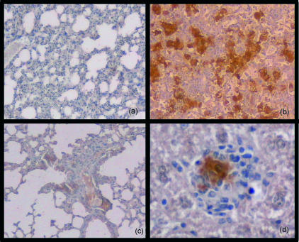
Figure 6.
Inducible nitric oxide synthase (iNOS) expression in lung tissue in mice during active, latent and reactivated tuberculosis infection. Mice were aerogenically infected with 30 colony-forming units (CFU) of Mycobacterium tuberculosis H37Rv, after which a group was treated with rifampicin and isoniazid (RMP-INH) for 8 weeks to induce latency of infection. Lung tissue of C57BL/6 mice was examined at 32 weeks postinfection. No iNOS staining was detected in mice during latent infection (a), whereas intense iNOS expression was observed in infected control mice (b). An augmented iNOS staining pattern was observed in mice 2 weeks after aminoguanidine-induced reactivation (c). Panel (d) shows a granuloma in the liver of an infected control mouse to illustrate the origin of iNOS protein from activated macrophages. Magnification: panels (a), (b) and (c) × 100; (d) × 400.

