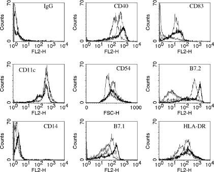
Figure 1.
Functional analysis of monocyte-derived DC. Day 5 monocyte-derived DC were incubated for a further 48 hr with DC media (thin-line) (medium-exposed DC), 1000 U/ml TNF-α (broken line) (TNF-α-exposed DC) or M. bovis BCG at a multiplicity of infection of 10 (bold line) (BCG-exposed DC) prior to staining with a panel of PE-conjugated antibodies for cell surface markers. Representative FACs plots of one donor are shown.

