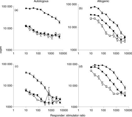
Figure 2.
Autologous (a,c) and allogeneic (b,d) mixed leucocyte reactions between media-exposed DC (○), TNF-α-exposed DC (▪); BCG-exposed DC (▴) and peripheral blood lymphocytes. Irradiated DC (stimulators) were cultured with autologous or allogenic PBL (responders) for 5 days. [3H]thymidine incorporation was determined during the last 18 hr of culture to measure T-cell proliferation. (a,b) show a representative PPD-responsive donor and (c,d) a PPD-non-responsive donor.

