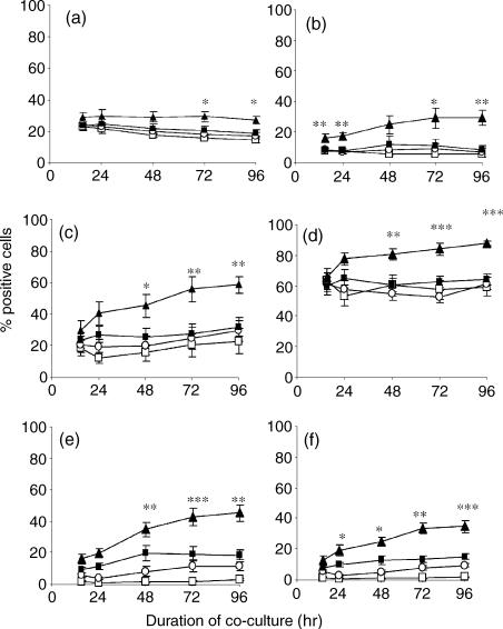
Figure 3.
Autologous peripheral blood lymphocytes were co-cultured with media (□), media-exposed DC (○), TNF-α-exposed DC (▪) or BCG-exposed DC (▴) at a ratio of 5 : 1 and the percentage of CD4+ (a,c,e) and CD8+ lymphocytes (b,d,f) expressing CD25 (a,b), CD54 (c,d), and CD71 (e,f) was determined by flow cytometry over a 96-hr period. The mean percentage of 10 donors is shown here. Any statistically significant increase of CD25, CD54, or CD71 on the surface of CD4 or CD8 T lymphocytes is indicated by *P < 0·05, **P < 0·01, ***P < 0·0001 (Student's t-test).

