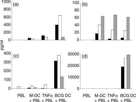
Figure 4.
Peripheral blood lymphocytes were co-cultured with autologous DC for 96 hr at a ratio of 5 : 1 and the supernatants were analysed for the presence of IL-2 (a), IL-4 (b), IL-10 (c) and IFN-γ (d) by solid-phase ELISAs. Three representative donors of 10 are shown (black, white and hatched bars). The lower levels of detection of the kits were 1·6, 1·6, 15·6 and 15·6 pg/ml for IL-2, IL-4, IL-10 and IFN-γ, respectively.

