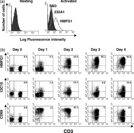
Figure 3.
Expression of MUC1 in activated T cells from a mixed lymphocyte reaction. (a) Responder peripheral blood mononuclear cells (PBMC), incubated for 6 days in the presence or absence of irradiated allogeneic stimulator cells, were stained with HMFG1–biotin, in association with CD3–fluorescein isothiocyanate (FITC) or CD25–FITC and analysed by flow cytometry. Numbers indicate the percentage of gated cells in the quadrant. (b) Cells activated with allogeneic PBMC every 7 days were stained for CD45RO and MUC1 (MF06 monoclonal antibody) and analysed by flow cytometry.

