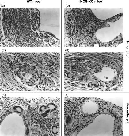
Figure 2.
Comparative histopathological investigation of Echinococcus multilocularis-infected wild-type (WT) mice (panels a, c and e) and inducible nitric oxide synthase (iNOS)-deficient mice (panels b, d and f) showed no difference between lesions of WT and iNOS-knockout (KO) mice at 1 and 4 months postinfection (p.i.). Panels (a), (b), (c) and (d) show lesions of mice at 1 month p.i. at low and high magnification. There was a massive fibrosis (panels a and b) with numerous multinucleated giant cells and epithelioid cells (panels c and d) in both wild-type (2a, 2c) and iNOS-KO mice (2b, 2d). There was still no significant difference between lesions of WT (Fig. 2e) and KO (Fig. 2f) mice at 4 months p.i. The magnification was ×160 for (a) and (b), and ×320 for (c), (d) and (f).

