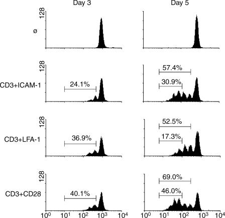
Figure 1.
Costimulation through ICAM-1 leads to a greater number of cells having divided three or more times compared to LFA-1. Peripheral blood T cells were labelled with CFSE and left non-stimulated or stimulated with antibodies against CD3 plus ICAM-1, LFA-1, or CD28. Cell division was assessed at day 3 (left panels) and day 5 (right panels) by dilution of CFSE. Percentages represent the total number of divided cells (left panels and top marker on the right panels) or the number of cells having divided three or more times (right panels, lower marker). Representative of five experiments.

