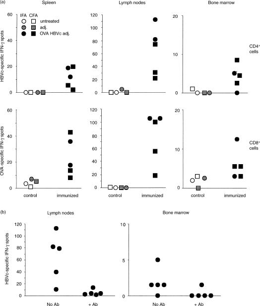
Figure 1.
IFN-γ ELISPOT analysis of antigen-specific CD4 and CD8 T cells from spleen, lymph nodes and bone marrow of OVA 257–264 and HBVc 128–140 peptide immunized and control B6 mice. (a)B6 female mice were immunized by two subcutaneous injections of OVA 257–264 and HBVc 128–140 peptides emulsified in either IFA or CFA, as indicated (Adj. = adjuvant). Two weeks after immunization, cells from spleen, inguinal and axillary lymph nodes and bone marrow of control and primed mice were purified and their response to each of the two immunizing peptides was analysed by IFN-γ ELISPOT. For the CD4 T-cell response, responder cells were stimulated with irradiated APC in the presence of either HBVc 128–140 peptide at 50 μg/ml or medium alone (P = 0·05 for bone marrow spots of immunized versus control mice). For the CD8 T-cell response, the responder cells were stimulated with either OVA 257–264 peptide pulsed or unpulsed irradiated APC in the presence of IL-2 at 20 U/ml. Responder spleen and lymph node cells were 250 000/well and bone marrow cells were 500 000/well. The HBVc 128–140 and OVA 257–264 peptide specific-IFN-γ spots of individual mice are represented, after subtraction of medium background. (b) B6 female mice were immunized by two subcutaneous injections of OVA 257–264 and HBVc 128–140 peptides emulsified in IFA and analyzed within 2 months after priming as in (a). Cells were incubated either in the presence or in the absence of the anti-I-Ab mAb AF6-120.1.2 in the culture medium during the 40-hr incubation of the ELISPOT test. The anti-I-Ab mAb AF6-120.1.2 was used as ascites at a 1:20 final dilution in the culture medium; at this dose, the IFN-γ ELISPOT response to the MHC class I-restricted OVA 257–264 peptide by lymph node cells from immunized mice was not blocked. The presence or absence of the anti-I-Ab mAb AF6-120.1.2 in the culture medium is indicated on the x-axis. The HBVc 128–140 peptide specific-IFN-γ spots of individual mice are represented, after subtraction of medium background (P < 0·05 for lymph node spots in the presence vs. the absence of the anti-I-Ab mAb).

